Case of the Week #564
Centro Médico Recoletas, Valladolid, Spain
Case Report: We present two cases at 20 and 27 weeks of pregnancy. In both cases, the mother's history was unremarkable. The ultrasound examinations performed in the first and second trimesters were normal. Although both cases seem to present the same finding, in reality they are distinct. We are looking for two answers that describe these two distinct findings. You may enter 2 answers for each guess.
Case 1
Case 2
View the Answer Hide the Answer
Answer
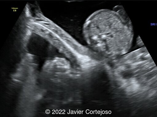
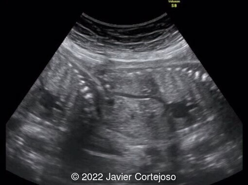
We present two types of ultrasound artifacts (mirror image and refraction artifact or double-image) in two distinct second and third trimester pregnancies. The first was pregnancy at 19+4 weeks gestational age with the fetus in cephalic presentation. Transabdominal ultrasound revealed an image suggesting the presence of an extrauterine gestational sac and containing fetal parts located laterally and behind the uterus. This image was obtained during certain moments of the scan, depending on the incidence angle of the ultrasound probe in the abdominal wall. The image of this second fetus is not as clear as the first one, making it impossible to assess its anatomy adequately. The movements of what appeared to be a second extrauterine fetus were synchronous with those of the intrauterine fetus, involving the same limbs, but in the opposite direction. In video 2 it is possible to observe that the extrauterine image is double.
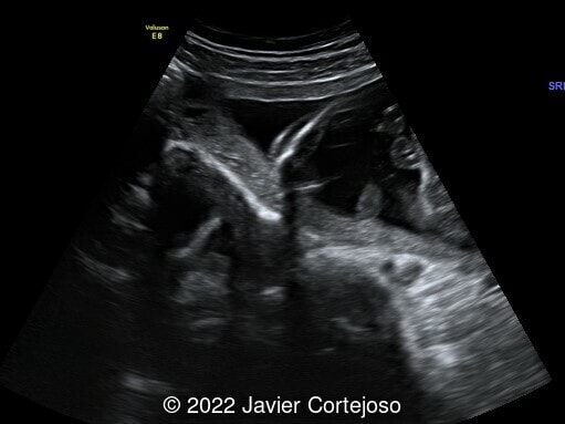
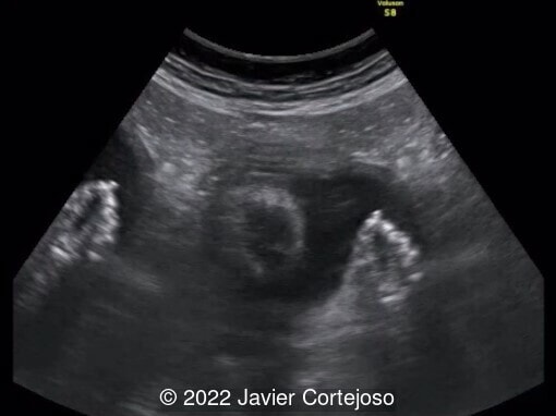
The second case corresponds to a gestation of 26+2 weeks with the fetus in breech presentation. As in the previous case, there is a duplication of different fetal parts (head, abdomen, thorax and an arm), but in this case the duplicated image has its same orientation and is not inverted.
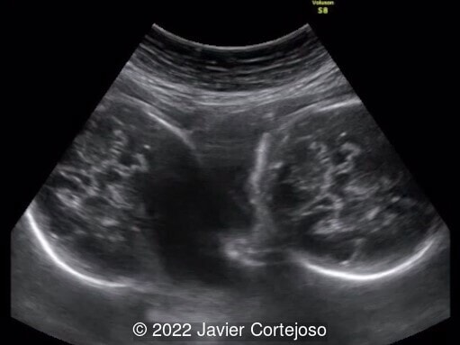
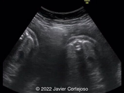
Fortunately, these artifacts are recognizable as the ghost image and usually depicts an anatomically impossible structure. In theory, the diagnostic possibilities that could be considered at first sight are twin pregnancy with two amniotic sacs or abdominal ectopic/heterotopic pregnancy. In the first case, the existence of a thick zone of separation between both possible gestational sacs, with a hyperreflective line inside, accompanied by the synchrony of inverted movements of both fetuses, made us suspect the presence of a mirror artifact. In the second, duplication of the image was confirmed by the "nodal point" in the outline of the anterior uterine wall and by the fact that when fetal movement is observed, both images move in unison. In both cases, presence of a single fetus was confirmed in subsequent scans and after the delivery of a normal newborn.
Discussion
In radiologic imaging, the term “artifact” is used to describe any part of an image that does not accurately represent the anatomic structures present within the subject being evaluated [1]. Ultrasound imaging artifacts of acoustic origin occur as displays that are duplicated (not real), are missing, or are of improper location, brightness, shape, or size [2].
Ultrasonography uses the physical properties of ultrasound waves to construct images of different tissues and structures. According to Kremkau [3], the premises for the design of ultrasound equipment are the following: (1) sound travels in straight lines, (2) echoes originate from objects located on the beam axis, (3) the amplitudes of returning echoes are related directly to the echogenicity of the objects that produced them, and (4) the distance to echogenic objects is proportional to the round-trip travel time. When any of these assumptions are violated, an artifact appears.
Ultrasound waves traveling through biologic tissues typically obey the laws of reflection and refraction. The most common image artifacts encountered in clinical practice can be grouped based on the following mechanisms:
-
The physics of reflection and refraction: reverberation, acoustic shadowing, mirror artifact, and refraction artifact
-
The properties of the ultrasound beam: side-lobe artifact and beam-width artifact
-
The equipment: near-field clutter [4].
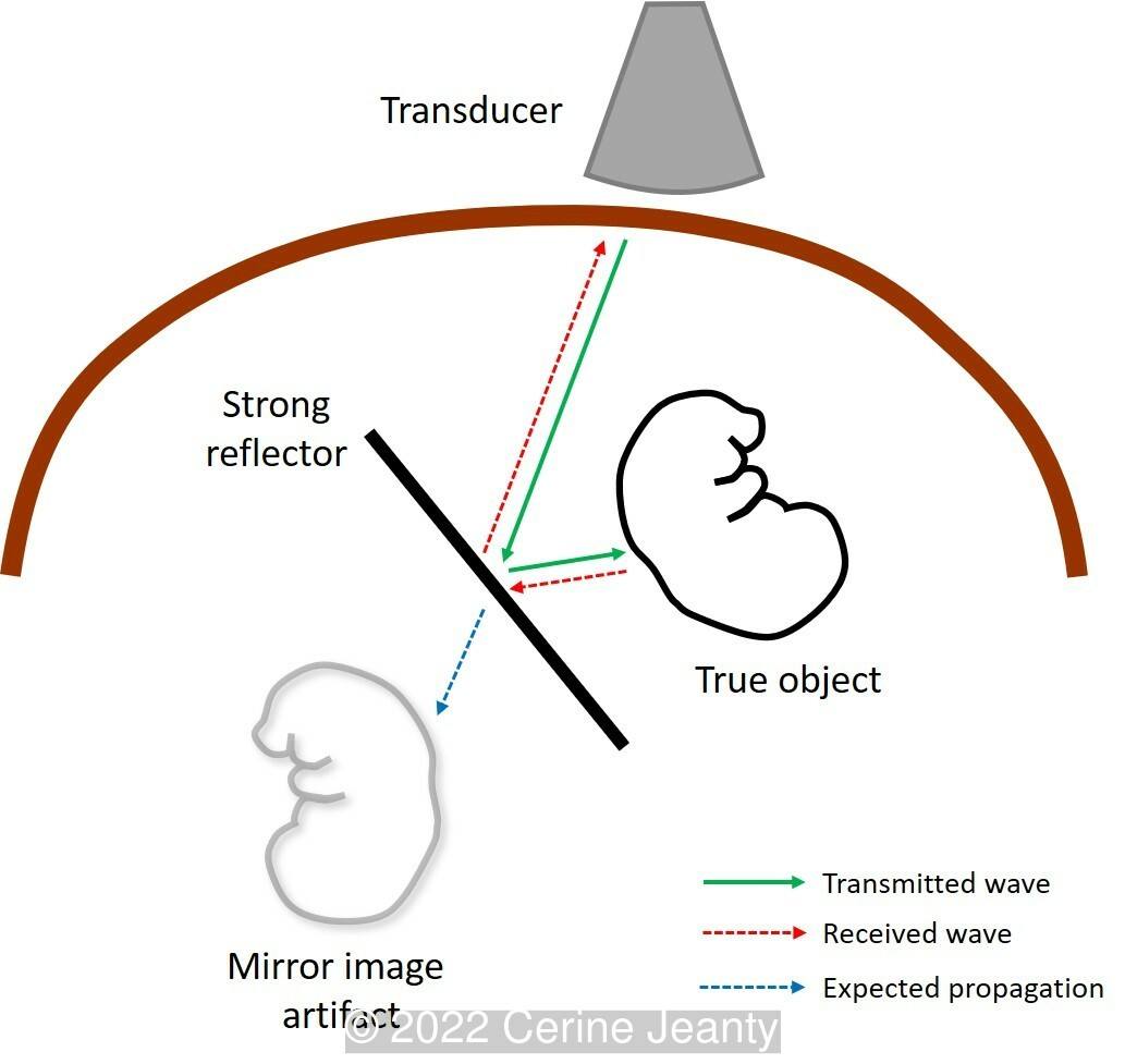
A mirror image artifact (Image 1) is created when the primary beam encounters a highly reflective surface (known as a “specular reflector”) that acts like a mirror [4]. Ultrasound waves reaching a strong reflector are reflected toward objects closer to the transducer than the reflector. These intervening objects reflect the waves back to the strong reflector, which in turn sends them back to the transducer. Because of the assumption of wave propagation (that all the returning sound waves comes from objects in the initial direction of the sound beam), the scanner displays these objects below the strong reflector, at a distance equal to the distance between strong reflector and the true intervening objects (Image 1). The false image acts as a mirrored object: inverted and moving in the opposite direction as the true structure. The existence of several reflectors could explain the appearance of multiple mirror images [5].
Mirror image artifact is commonly seen in the liver leading to duplication of hepatic structures caused when the diaphragm acts as a strong reflector [6]. The typical display of the mirror artifact consists of two similar structures separated and at similar distances from the reflective interface. The mirror image is hypoechoic, blurred, and distorted compared to the image of the real structure. The image found in the “phantom” uterus may be somewhat different from the intrauterine image. This is because the mirror is not exactly perpendicular to the plane of the probe, so the uterus is being imaged in a slightly different cross-section [5].
It has been reported in vascular, abdominal, cardiac, and musculoskeletal ultrasound, but is uncommon in obstetric ultrasound, having been published on seven occasions to the best of our knowledge, and practically always in the first trimester (with transabdominal and transvaginal probes) [5, 7-10]. There is only one case published in the second trimester of pregnancy [11]. In the first trimester, the psoas muscle acts as a reflective surface when the bladder is over distended and the uterus is pushed up into the abdomen [8]. At more advanced gestational age, gas and fluid in the bowel are the main contributors to the ultrasound artifact, not only creating a strong interface with the posterior uterine wall, but also serving as a space of representation of the mirrored image in the ultrasound screen [11].
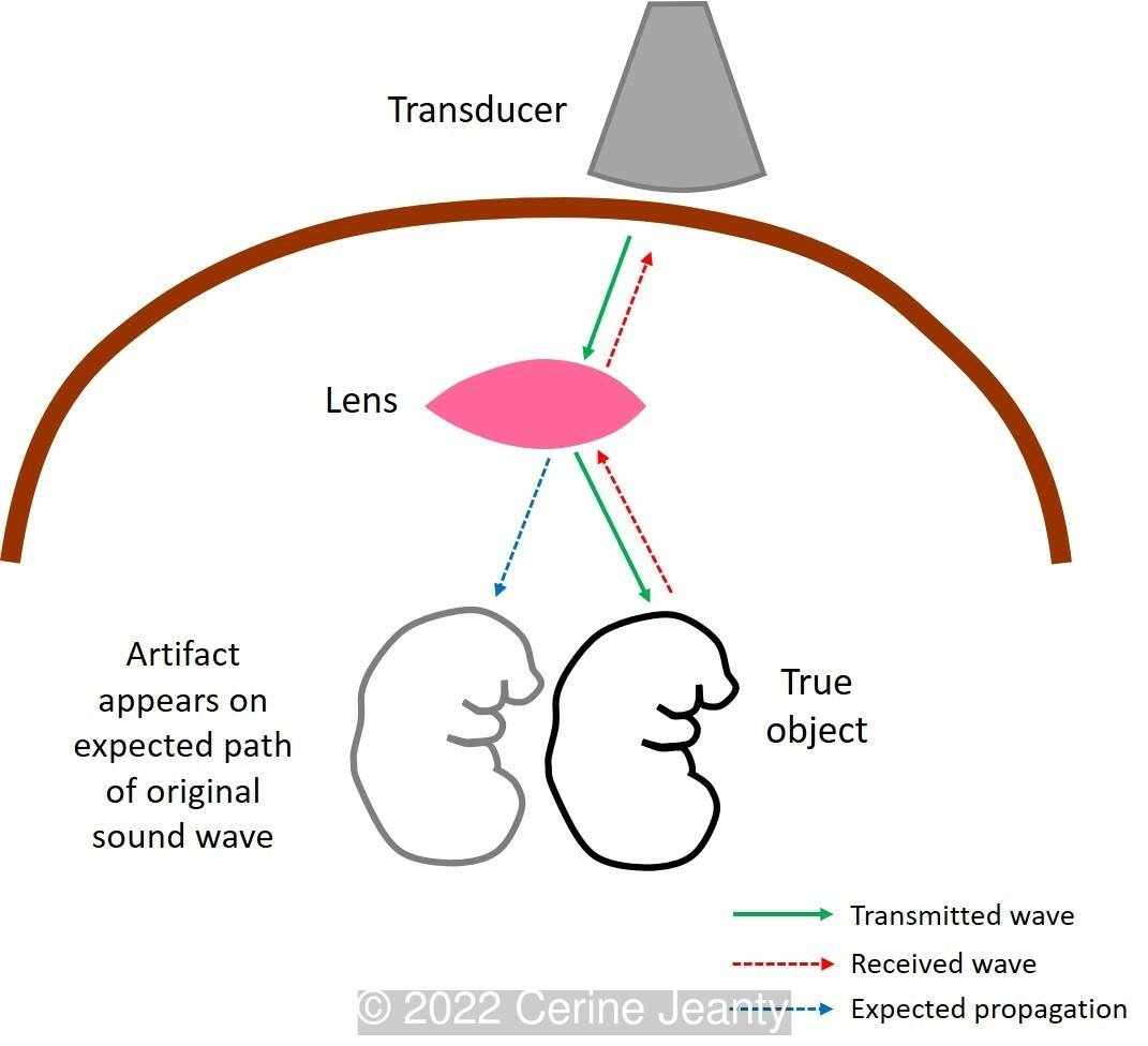
A refraction artifact (Image 2) is produced when the probe is placed in the abdominal midline including the lateral margins of the rectus muscles [12,13]. Refraction can result in improper positioning of an object on the display screen or it may cause a single real structure to appear as two objects due to the existence of a refracting structure close to the transducer, which behaves like a lens [3]. Ultrasound waves directed through the ‘‘lens’’ are refracted toward the object and then re-refracted back to the original direction of transmission on the return acoustic path, resulting in a duplicate image of this object but in the original direction of the beam (Image 2) [4]. The rectus muscle interposed between the transducer and the area of interest acts like a lens and refracts the ultrasound beam, producing the duplication (or even triplication) of a single object [13]. When a part of the image of a relatively spherical structure such as the pregnant uterus is displaced by the refraction caused by the edge of the rectus muscle, the overlapping image produces a discontinuity (“nodal point”) in the uterine outline, and the shadow of the edge effect is evident [12]. Buttery and Davison call it the “ghost artifact” [12], although Müller et al prefer to call it "double image artifact" [13], which clearly describes the finding and avoids introducing confusing terms. Ahn et al speak of a ghost twin as a consequence of a mirror artifact, leading to further confusion in using this "ghost" terminology [11].
Although the duplication artifact is more commonly observed in the pelvis, it may also be seen in the upper abdomen. At first glance, it might look like a mirror artifact. However, we can differentiate them if we take into account that in the refraction artifact the repeated image appears next to instead of underneath the true image, and is not reversed as in the mirror artifact [3]. Also, when fetal movement is seen on real-time ultrasound examination, the two images move in unison [12].
Ultrasound artifacts are created by the machine’s interpretation of returning echoes but do not correspond to the actual anatomy of the patient. Physicians have to be aware of mirror or refraction artifact to avoid a misdiagnosis of heterotopic or twin pregnancy. Once identified, these artifacts can be decreased or eliminated by moving the transducer to center it on the region of interest or by placing the focal zone at the level of interest in the real structure [6, 14]. Maneuvers (transabdominal-transvaginal probe change, filling or emptying the bladder, maternal position changes) to verify correct diagnosis and resolve potential mirror or double image artifacts should be used before proceeding with surgical management of a false diagnosis of heterotopic pregnancy [9]. In the same way, more detailed imaging techniques such as magnetic resonance imaging (MRI) may be considered [10].
References
[1] Feldman MK, Katyal S, Blackwood MS. US artifacts. Radiographics. 2009 Jul-Aug;29(4):1179-89.
[2] Kremkau FW, Taylor KJ. Artifacts in ultrasound imaging. J Ultrasound Med. 1986 Apr;5(4):227-37.
[3] Kremkau FW. Artifacts. In: Sonography Principles and Instruments, ninth edition. Elsevier, St Louis, MO, 2016; pages 183-216.
[4] Bertrand PB, Levine RA, Isselbacher EM, et al. Fact or Artifact in Two-Dimensional Echocardiography: Avoiding Misdiagnosis and Missed Diagnosis. J Am Soc Echocardiogr. 2016 May;29(5):381-91.
[5] Lim BH, Amos M, Fairhead AC. The mirror image artifact of early pregnancy. Ultrasound Obstet Gynecol. 2003 May;21(5):518-20.
[6] Khalili K, Yu H, Jesurum A, and Levine D. Ultrasound Artifacts: A Virtual Chapter. In: Rumack CM and Levine D, ed. Diagnostic Ultrasound, 5th ed. Elsevier, Inc. Philadelphia, PA, USA, 2018; pages e1-e38.
[7] Miglietta F, D'Antonio F, Matarrelli B, et al. Mirror-image artifact of early pregnancy on transvaginal sonography. J Ultrasound Med. 2012 Nov;31(11):1858-9.
[8] Malhotra R, Bramante RM, Radomski M, et al. Mirror image artifact mimicking heterotopic pregnancy on transvaginal ultrasound: case series. West J Emerg Med. 2014 Sep;15(6):712-4.
[9] Ahmed R, Samardzic D, Santos MA, et al. Just a mirage: heterotopic intrauterine and twin ectopic pregnancy mimicked by mirror imaging on ultrasound. Radiol Case Rep. 2017 Apr 8;12(2):422-426.
[10] Russell J, Paterson K, Cooper S. Apparent Heterotopic Pregnancy Created by Mirror Image Artifact. J Obstet Gynaecol Can. 2017 Nov;39(11):961-962.
[11] Ahn H, Hernández-Andrade E, Romero R, et al. Mirror artifacts in obstetric ultrasound: case presentation of a ghost twin during the second-trimester ultrasound scan. Fetal Diagn Ther. 2013;34(4):248-52.
[12] Buttery B, Davison G. The ghost artifact. J Ultrasound Med. 1984 Feb;3(2):49-52.
[13] Müller N, Cooperberg PL, Rowley VA, et al. Ultrasonic refraction by the rectus abdominis muscles: the double image artifact. J Ultrasound Med. 1984 Nov;3(11):515-9.
[14] Quien MM, Saric M. Ultrasound imaging artifacts: How to recognize them and how to avoid them. Echocardiography. 2018 Sep;35(9):1388-1401.
Discussion Board
Winners

Danilo Feitosa Brazil Physician

Fatih ULUC Turkey Physician

Igor Yarchuk United States Sonographer

Dmitry Abelov Russian Federation Physician

Ana Ferrero Spain Physician

Tatiana Koipish Belarus Physician

Rushina Patel United States Sonographer
Rebecca Evans Australia Sonographer

Shari Morgan United States Sonographer

ALBANA CEREKJA Italy Physician

Murat Cagan Turkey Physician

TEJAS TAMHANE India Physician

maria maxfield Australia Sonographer