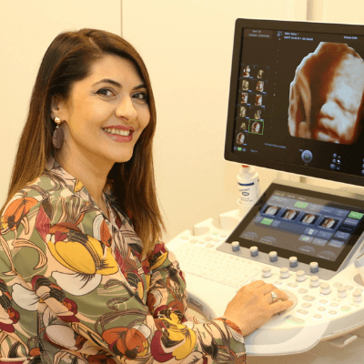
Case of the Week #566
Yaroslavl, Russia
Reviewer: Javier Cortejoso
Case Report: A 23-year-old G1P0 with no remarkable medical history was scanned at our unit at 18 weeks of gestation. Ultrasound examination at the first screening and at 17 weeks were reported to be normal.
View the Answer Hide the Answer
Answer
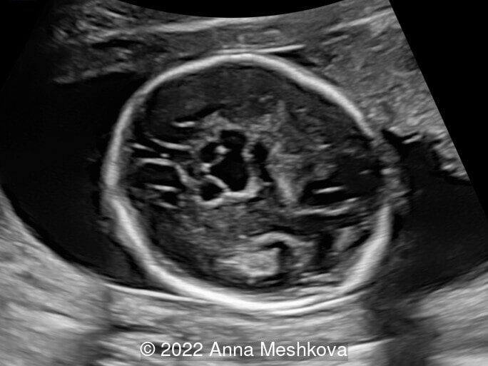
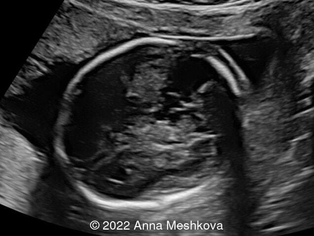
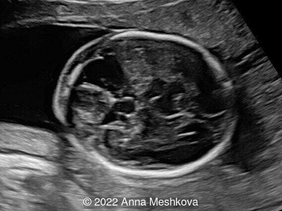
We present a case of intracranial teratoma.
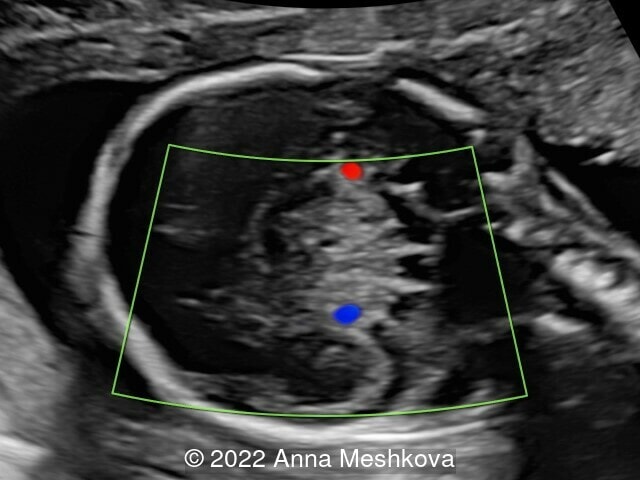
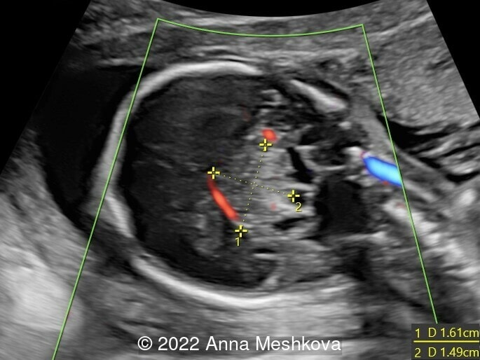
Ultrasound imaging revealed the lesion approximately 26mm x 17mm x 23 mm located in the middle of the brain. The solid hyperechoic component measured 15mm x 16 mm. It was well defined, rounded, echogenic and heterogeneous with spots of hyperechogenicity. Color Doppler examination showed the presence of vascularization in the lesion. The other cerebral structures had a normal appearance, but some of them were compressed and displaced by the lesion. There was no extracranial abnormality.
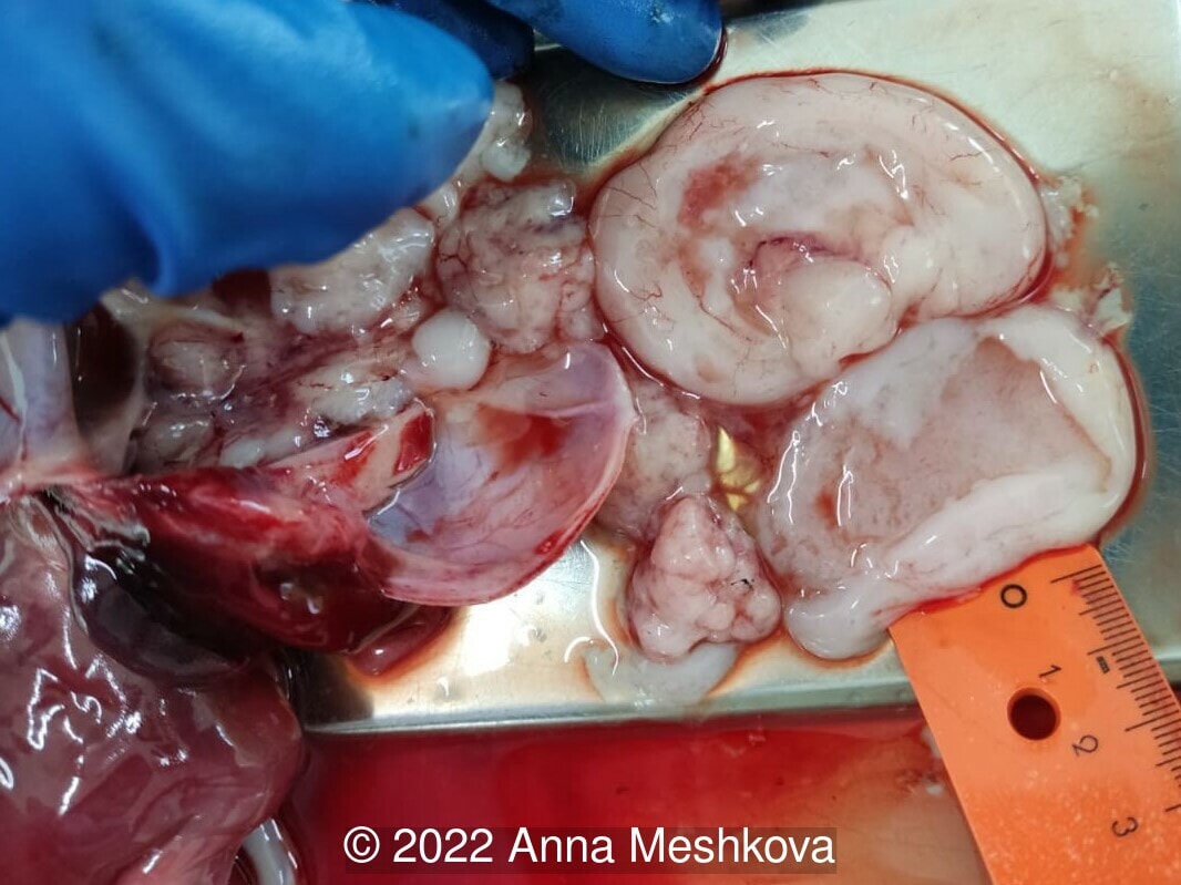
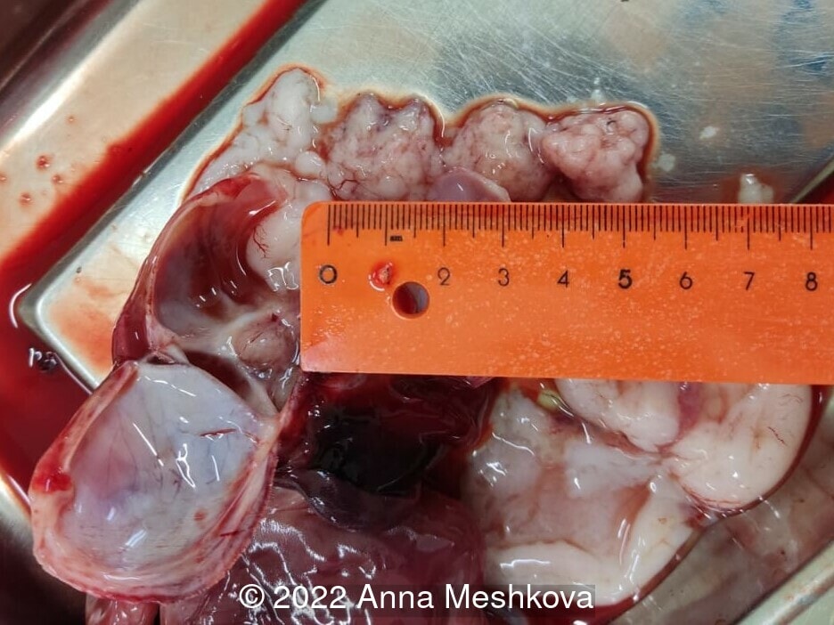
The lesion was suspected to be a cerebral tumor due to its solid features and the presence of vascularization. Differential diagnoses were cerebral hemorrhage. Because of the early appearance of the lesion and its significant size, the prognosis was considered to be poor. A termination of pregnancy was discussed with the parents and was carried out at 19 weeks of gestation. Postmortem examination confirmed the diagnosis of intracranial teratoma, containing lung, bone, cartilage and neuroectoderm, with destruction of the central and right cerebral structures. Also the structures of the posterior fossa were displayed toward the cisterna magna.
Discussion
Congenital intracranial tumors are a rare occurrence, accounting for only 0.5–1.5% of all childhood brain tumors [1]. The most common of these tumors are teratomas, which comprise between 29% and 50% of central nervous system tumors. In childhood, teratomas have two peaks in their age distribution: 10% occur before 5 years of age whereas 48% occur between 5 and 14 years of age. Some consider that the incidence of teratoma in the first peak may be related to the sequestration of blastocyst cells before differentiation has occurred [2].
Teratomas are considered to be a subtype of germ cell tumors, containing all types of embryonic germ cell layers: ectoderm, mesoderm, and endoderm [3]. They are often immature because of primitive neural elements and, rarely, a component of mixed malignant germ cell tumors. Although they are tumors of the gonads, they can originate outside of the gonads. Extragonadal teratomas are congenital midline tumors, located both intracranially and extracranially, such as in the mediastinum and retroperitoneum. They are considered to originate from primordial germ cells that fail to migrate properly during the initial weeks of embryonic development [4].
Congenital intracranial teratomas are the most frequent intracranial tumor of the perinatal and neonatal period, comprising around 62% of prenatally detected cases [5]. They occur five times more frequently than astrocytoma, which is the second most common type of congenital intracranial tumor [6,7]. Teratomas occur almost exclusively above the tentorium. In a review by Wakai et al of 200 cases, only one was found in the posterior fossa [8].
Prenatal diagnosis of these tumors has increased in recent years. Diagnosis can be suspected on routine fetal ultrasound examination. It may show intrauterine ventriculomegaly, enlarged biparietal diameter, and/or brain tumor [9]. Intracranial teratomas are usually diagnosed by sonographic examination in the second or third trimester and early diagnoses are not as common [10-16]. If fetal ultrasound is suggestive of ventriculomegaly due to a tumor, MRI is advised for further localization, size estimation, and determination of the tumor extent, as well as ventriculomegaly [9,17]. Karyotyping is not recommended routinely but should be discussed if other abnormalities are present [18].
Subdural hemorrhage should be considered as a differential diagnosis in cases of intracranial space-occupying lesion [19]. Like teratomas, it is usually located in the supratentorial region. Predisposing factors may include maternal anticoagulant therapy, abdominal trauma, and fetal coagulation disorders, although may also occur spontaneously. In the present case, color Doppler imaging showed vascularization in the lesion which helped confirm the diagnosis of tumor.
Early diagnosis is important as termination of pregnancy may avoid obstetric complications. Ventriculomegaly may be responsible for macrocephaly and secondary dystocia by cephalopelvic disproportion. In this situation, Cesarean section is performed in about 60% of cases [5,9,17,20-21]. In addition to hydrocephalus, other common clinical conditions at the time of presentation are polyhydramnios, followed by respiratory distress and stillbirth [20].
The prognosis for intracranial teratomas is poor and the survival rate is less than 10% [5,14,22,23]. A large number of affected fetuses die prenatally and the rest shortly after birth [6]. Outcomes are dependent on the time of diagnosis and the size of the teratoma. In a review of 73 teratomas, the 1-year survival rate of infants with congenital intracranial teratoma was 7.2% [8]. Fukuoka et al describe an infant that survived until 3 years of age with adjuvant chemotherapy and complete surgical resection [24]. Partial resection does not appear to confer same benefits on long-term survival [25]. The location and the size of the teratoma are considered as significant prognostic factors rather than the histological grade [6], with the rarer posterior fossa teratomas being associated with worse outcomes.
References
[1] Buetow PC, Smirniotopoulos JG, Done S. Congenital brain tumors: a review of 45 cases. AJNR Am J Neuroradiol. 1990;11(4):793–799.
[2] Sano K. Intracranial dysembryogenetic tumors: pathogenesis and their order of malignancy. Neurosurg Rev 2001; 24: 162– 167; discussion 168–170.
[3] Algahtani HA, Al-Rabia MW, Al-Maghrabi HQ, et al. Posterior fossa teratoma. Neurosciences. 2013;18(4):371–374.
[4] Slavin KV, Ausman JI. Tumors of the pineal region. In: Rengachary SS, Wilkins RH, editors. Principles of neurosurgery. London: Mosby; 1994. pp. 29.1–29.16.
[5] Schlembach D, Bornemann A, Rupprecht T, et al. Fetal intracranial tumors detected by ultrasound: a report of two cases and review of the literature. Ultrasound Obstet Gynecol 1999; 14: 407– 418.
[6] Isaacs H., Jr Perinatal (fetal and neonatal) germ cell tumors. J Pediatr Surg. 2004;39(7):1003–1013.
[7] Raisanen JM, Davis RL. Congenital brain tumors. Pathology. 1993;2(1):103–116.
[8] Wakai S, Arai T, Nagai M. Congenital brain tumors. Surg Neurol. 1984;21:597–609.
[9] Ferreira O. Prenatal diagnosis of intracranial tumors: a case report. Pediatr Neurosurg. 1993;19:84–88.
[10] Horton D, Pilling DW. Early antenatal ultrasound diagnosis of fetal intracranial teratoma. Br J Radiol 1997; 70: 1299– 1301.
[11] Arslan E, Usul H, Baykal S, et al. Massive congenital intracranial immature teratoma of the lateral ventricle with retro-orbital extension: a case report and review of the literature. Pediatr Neurosurg 2007; 43: 338– 442.
[12] Mazouni C, Porcu-Buisson G, Girard N, et al. Intrauterine brain teratoma: a case report of imaging (US, MRI) with neuropathologic correlations. Prenat Diagn 2003; 23: 104– 107.
[13] Muhonen MG, Bierman JS, Hussain NS, et al. Giant intracranial teratoma and lack of cortical development in a fetus. Case report. J Neurosurg 2005; 103 (Suppl): 180– 183.
[14] Woodward PJ, Sohaey R, Kennedy A, et al. From the archives of the AFIP: a comprehensive review of fetal tumors with pathologic correlation. Radiographics 2005; 25: 215– 242.
[15] Rickert CH, Probst-Cousin S, Louwen F, et al. Congenital immature teratoma of the fetal brain. Childs Nerv Syst 1997; 13: 556– 559.
[16] Saada J, Enza-Razavi F, Delahaye S, et al. Early second-trimester diagnosis of intracranial teratoma. Ultrasound Obstet Gynecol. 2009 Jan;33(1):109-11
[17] Hoff NR, Mackay IM. Prenatal ultrasound diagnosis of intracranial teratoma. JCU. 1980;8:247–249.
[18] Schwartz S, Raffel LJ, Sun CC, et al. An unusual mosaic karyotype detected through prenatal diagnosis with duplication of 1q and 19p and associated teratoma development. Teratology 1992; 46: 399– 404.
[19] Meagher SE, Walker SP, Choong S. Mid-trimester fetal subdural hemorrhage: prenatal diagnosis. Ultrasound Obstet Gynecol 2002; 20: 296– 298.
[20] Takaku A, Kodama N, Ohara H, et al. Brain tumor in newborn babies. Childs Brain. 1978;4(6):365–375.
[21] Cavalheiro S, Moron AF, Hisaba W, et al. Fetal brain tumors. Childs Nerv Syst. 2003;19:529–536.
[22] Pinto V, Meo F, Loiudice L, D et al. Prenatal sonographic imaging of an immature intracranial teratoma. Fetal Diagn Ther 1999; 14: 220– 222.
[23] Chien YH, Tsao PN, Lee WT, et al. Congenital intracranial teratoma. Pediatr Neurol 2000; 22: 72– 74.
[24] Fukuoka K, Yanagisawa T, Suzuki T, et al. Successful treatment of hemorrhagic congenital intracranial immature teratoma with neoadjuvant chemotherapy and surgery. J Neurosurg Pediatr. 2014;13(1):38–41.
[25] Robles Fradejas M, Gonzalo Garcia I, De Las Casas Quispe AC, et al. Fetal intracranial immature teratoma: presentation of a case and a systematic review of the literature. J Matern Fetal Neonatal Med. 2017;30(10):1139–1146.
Discussion Board
Winners

Dianna Heidinger United States Sonographer
Danilo Feitosa Brazil Physician

Fatih ULUC Turkey Physician

Umber Agarwal United Kingdom Maternal Fetal Medicine

Andrii Averianov Ukraine Physician

Ana Ferrero Spain Physician

Mayank Chowdhury India Physician

Adrian Popa Romania Physician
DAVID BEAUMONT United Kingdom Physician

Ivan Ivanov Russian Federation Physician
Halil Mesut Turkey Physician

Miğraci Tosun Turkey Physician

Rushina Patel United States Sonographer

Anita Silber Israel Physician

lan nguyen xuan Viet Nam Physician
Sahar Salama United States

Suat İnce Turkey Physician
santosh shahare United States Physician

Muradiye Yıldırım Turkey Physician
Umutcan KAYIKÇI Turkey Physician
Rasha Abo Almagd Egypt Physician

José Lambertino Colombia Physician
Halil Korkut Dağlar United States Physician

Prakriti Patil India Physician
KHALED RAMADAN Egypt Physician
Seda Cam Turkey Physician
Almaz Kinzyabulatov Russian Federation Physician
Sushant Jain United States Medical Student