Case of the Week (COW)

Current Case of the Week (COW)

Current Case of the Week (COW)
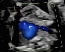
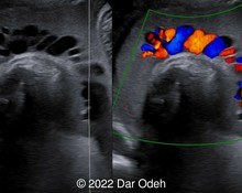
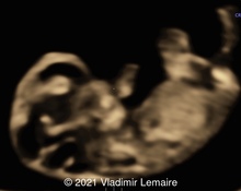
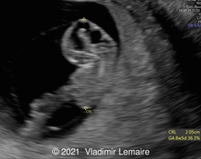
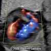
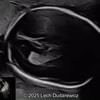
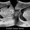
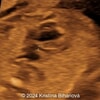
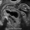
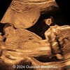
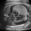
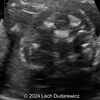
A 27-year-old G1P0 woman presented to our maternal fetal medicine unit at 26 weeks, 4 days of gestation for a growth scan. The fetus was female with low-risk noninvasive prenatal testing. The following findings were observed.
Submit Your Answer
To join the community and participate in solving current cases, simply click the "view current case" button below. This will take you to the case summary page with full details and additional media. To participate, you will first need to create an account or sign-in. You can then submit your answers.
You can only submit your answers once, but you get three answers. The correct answer is revealed at the end of the posting period.
Physicians
Previous Winners
-

Javier Cortejoso, Spain
Cases Solved: 4 -
Tetiana Ishchenko, Ukraine
Cases Solved: 4 -
Alexandr Krasnov, Ukraine
Cases Solved: 4 -
Ionut Valcea, Romania
Cases Solved: 4 -
Ivan Ivanov, Russian Federation
Cases Solved: 3
First-Time Winners
-

Ali Ozgur Ersoy, Turkey
Cases Solved: 3 -
Jagdish Suthar, India
Cases Solved: 2 -
Eylem Eşsizoğlu, Turkey
Cases Solved: 2 -
Zhanna Bondarchuk, Ukraine
Cases Solved: 2 -
Petra Tallova, Slovakia
Cases Solved: 2
Sonographers
Previous Winners
-

Dianna Heidinger, United States
Cases Solved: 4 -
Kimberly Delaney, United States
Cases Solved: 3 -
Anette Beverdam, Netherlands
Cases Solved: 2 -
Igor Yarchuk, United States
Cases Solved: 1 -
Rebecca Evans, Australia
Cases Solved: 1
First-Time Winners
-

Fred Pop, Uganda
Cases Solved: 3 -
Joanne Maloney, United States
Cases Solved: 2 -
CHEN YANG, China
Cases Solved: 2 -
ALPESH PANCHOLI, United States
Cases Solved: 2 -
Oscar Hernández, United States
Cases Solved: 1
Contributors
Top Contributors
-

Lech Dudarewicz, Wallis and Futuna
Article & Case Contributions: 3 -
Tushar verma, India
Article & Case Contributions: 1 -
Javier Cortejoso, Spain
Article & Case Contributions: 1 -
Vladimir Lemaire, United States
Article & Case Contributions: 1
Previous winners: Users who have been recognized on a "Top Winners list" in years past; First-time winners: Users who have not yet been on the annual "Top Winners list." The Sonographers category also incorporates "other" job titles.

News & Notes
Dear Esteemed Users of TheFetus.net
This year there were 363 participants who responded correctly to at least one Case of the Week. Congratulations to everyone who solved these challenging cases!
We have finalized the annual winner's list that recognizes those who have persistently found the correct diagnosis to the Case of the Week. Congratulations! If I have made a mistake or you would like to fill in your bio or profile picture, please email me: cow@thefetus.net
I… read the full entry
TheFetus.net

